The History of Ultrasound and How It Changed Varicose Vein Treatment
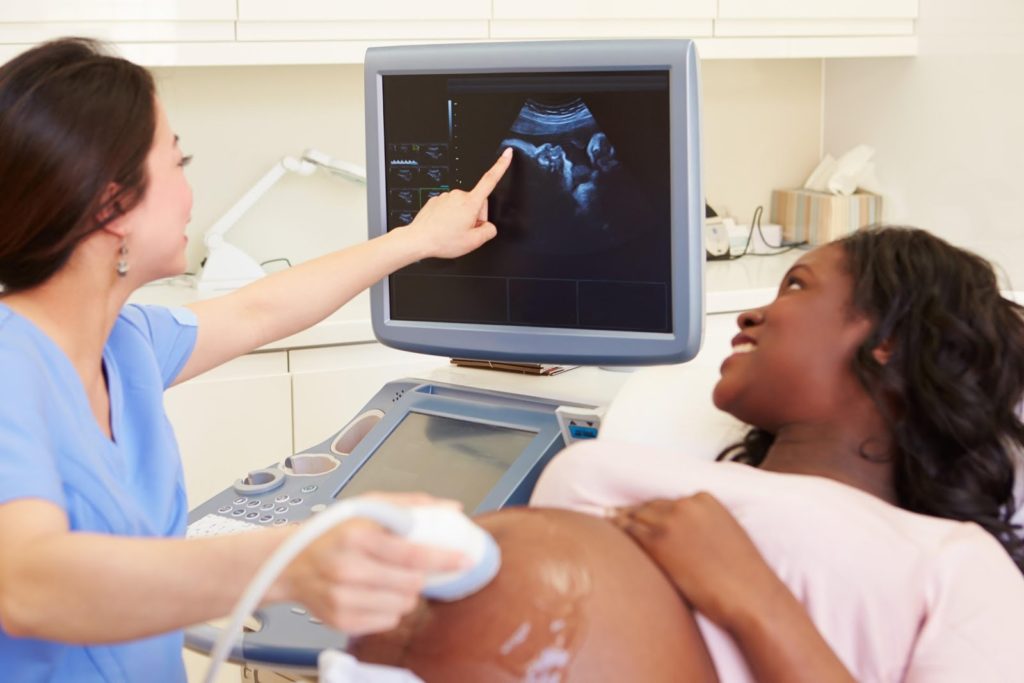
Like so much of our medical advancements, they seem so commonplace today that we hardly blink an eye. Most people associate ultrasound technology with prenatal care—seeing that cute little baby for the first time. The technology of ultrasound, however, extends into other areas of the medical field. It has helped different treatments take great strides and improve diagnoses. Today, ultrasound is used for the treatment of varicose veins and facilitated the assessment of vein health without an invasive procedure. Let’s take a look at how this technology got started and how it made its way to venous disease treatment clinics.
It Begins With Bats and Their Amazing Echolocations
We’ve all heard that common expression of “blind as a bat.” As it turns out, bats are pretty blind; sight is not exactly their strongest sense. So how do these flying creatures get around without seeing where they’re going? Echolocation is the active use of sonar along with physical features and other physiological adaptations has given bats the ability to “see” with sound. It is the concept of this ability that forms the basis for what we know today as ultrasound. Bat calls are categorized according to frequency, duration, and intensity and they fall beyond the range of human hearing. They emit ultrasonic waves with high frequency. It is with the concept of capturing ultrasound that we are able to create an apparatus that uses these frequencies to see beneath human tissue.
Scientists and medical doctors have always sought the ability to see through the body. Around 1877, French physicist Pierre Curie’s discovery of piezoelectricity. This was, for many, the beginning of the ultrasound technology as we would come to know it today. Later, sonographic imaging was developed by another Frenchman Paul Langevin.
X-Rays — A Precursor of Ultrasound
In 1865, William Conrad Roentgen discovered x-ray technology when working with a cathode-ray tube in his laboratory. The ray that he discovered went through the thick paper and he then discovered that it could also trespass human tissue. As these rays were discovered, the scientific community was ecstatic and the public too was fascinated about this exciting new discovery that allowed doctors to see through human skin. Imagine that! It was pretty groundbreaking stuff. Scientists from different fields dropped what they were doing and continued investigating and researching these mysterious rays.
The conscious search for ultrasonic technology was largely due to the first World War. The French government called Langevin to help with the detection of submerged submarines underwater. And while he barely missed the mark, Langevin did contribute to the overall basis of sonar detection that would become so crucial in proceeding conflicts like World War II.
Ultrasound for Medical Purposes
It wasn’t until the 1920s and 1930s that ultrasound was utilized for medical purposes. Some of the first people to benefit from it were European soccer players who had ultrasound used for physical therapy. It was also used for sterilization of vaccines and for cancer therapy in combination with radiation therapy. In the 40s, ultrasound was used for arthritic pains to gastric ulcers to even skin conditions.
With the passing years, other pioneers got in on the advancement of ultrasound. In 1950, ultrasound was used for detecting tumors in the brain. Two men by the name of John Reid and John Wild would come to build a linear, handheld, B-mode instrument for breast tumors.
Ultrasound for Venous Disease Treatment
For doctors who specialize in the treatment of venous disease including varicose veins, ultrasound has allowed them to detect these damaged veins and assess the problem thoroughly with a quick and non-invasive exam. Venous ultrasound works by taking images using sound waves. Not unlike the way that bats can ‘see’ through the darkness by bouncing off sounds, ultrasound will identify images using high-frequency sound waves. The ultrasound machine transmits high-energy sound pulses into your body. These pulses travel into the body and bounce off internal tissues and create a type of echo. These ‘echoes’ or reflected waves are picked up by the probe and transmitted back to the machine.
Ultrasound is used to evaluate your vein function and check for blood clots. The technique is used to look at all deep and superficial veins in the legs. The veins are checked on their starting point and endpoint. Another condition such as deep vein thrombosis is also diagnosed and found through ultrasound. This condition is found through a dual process called Duplex Ultrasound and is so successful in detecting DVT in the large veins above the knee. This technique is said to detect 95 percent of deep vein thrombosis. Ultrasound is also used to detect this condition throughout the leg.
Get Accurate Diagnosis with El Paso Varicose Veins Laser Clinic
Here at El Paso Varicose Vein Laser Clinic, we use today’s latest technology and techniques to diagnose, treat, and monitor the venous disease, varicose veins, and deep vein thrombosis. Varicose veins can be a painful condition that prevents people from living the active lifestyle they seek. If you think you might suffer from varicose veins, call the clinic today and come in for an ultrasound.